


This disease is poorly understood, although several thousand observations with diagnosis and subsequent treatment have been described.
The multitude of diversity and non-specificity of the clinical picture of varicose veins of the small pelvis lead to gross errors on the part of the diagnosis, which in the future affects the consequences.
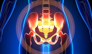
Characteristics of varicose veins of the small pelvis
The veins of the pelvis are several times longer than the arteries, which leads to their greater capacity. This is due to the phylogenesis of the vascular system of the pelvic region. The pelvic veins are highly adaptable and potentially prone to remodeling, which contributes to the formation of a densely interwoven network.
The speed and direction of blood flow are regulated by valves, which are controlled by complex humoral mechanisms. The valves balance the pressure in different parts of the venous network.
When the valves cease to perform their functions, blood stagnation develops, this leads to vascular pathology and the formation of varicose veins. The uniqueness of the pelvic veins lies in the fact that the wide ligaments of the uterus, which keep the lumen of the vessel wide, can narrow it, causing pathology.
Causes of occurrence
Pathological pelvic venous dilatation may be due to the following reasons:
- Disruption of blood outflow tract;
- Obliteration of the vein trunk;
- Compression of collateral trunks by an altered position of the uterus, for example, in retroflection;
- ovarian vein valve insufficiency (congenital or acquired);
- Obstructive postphlebitic syndrome;
- Connective tissue pathology;
- Arteriovenous angiodysplasia;
- Prolonged sitting, hard physical labor;
- Varicose veins of the lower extremities;
- Pregnancy (3 or more) and childbirth (2 or more);
- Diseases of the female genital area (chronic salpingo-oophoritis, ovarian tumors, uterine fibroids and genital endometriosis);
- Adhesion of the pelvic organs;
- Obesity.
Classification by disease grade
By the size of the dilated vein, the following degrees are distinguished:
- up to 0. 5 cm, "corkscrew" course of vessels;
- 0. 6-1 cm;
- more than 1 cm.
Variants of the course of the disease
- varicose veins of the perineum and the vestibule of the vagina;
- syndrome of venous congestion of the small pelvis;
Symptoms
- The most common - frequent pains in the lower abdomen, perineum after long static and dynamic overstrain. The pain increases in the second phase of the cycle, after hypothermia, fatigue, stress, exacerbations of various diseases.
- Feeling "out of place", pain during and after sex.
- Dysmenorrhea - menstrual irregularities, including pain.
- Secretion, more than normal, of the glands of the genital tract.
- Blood stagnation leads to infertility, miscarriage, abortion.
- Violation of urination due to the expansion of the veins of the bladder.
Diagnostics
Diagnosis of the disease only by complaints is successful only in 10% of cases.
Palpation of the inner walls of the pelvis, makes it possible to feel the oblong seals and venous nodes. When viewed in the mirrors, cyanosis of the vaginal mucosa is visible.
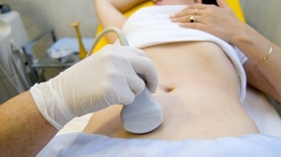
The procedure of choice is an ultrasound examination with color Doppler mapping, which allows to detect not only varicose ovarian veins, but also venous thrombosis, post-thrombophlebitic occlusions. Ultrasound shows tortuosity, "worm-like", structures without signal reflection, localized on the lateral surface of the uterus.
The Doppler effect is based on blue and red tint of venous and arterial blood flow, respectively.
The device for ultrasound examination with the help of a special program recognizes the movement of blood from the sensor and in the other direction, calculates the blood flow velocity and the type of vessel.
But the exact definition of a vein or an artery remains with the doctor. The Doppler method works in almost all cases, exceptions to the rules are dictated by our body, since the blood that flows from the heart is not always arterial and vice versa.
Thus, the doctor of ultrasound diagnostics sees this arterial or venous vessel, its size, blood flow rate in it and many indicators that are not needed by an ordinary person, but play an important role in making a diagnosis. For this, transabdominal and transvaginal sensors are used.
In 5. 7% of cases, the disease is recognized by chance at screening. Normally the diameter of the vena ovarica is 0. 4 cm.
CT and MRI are highly accurate. With these methods, it is possible to detect accumulations of varicose veins in the ligaments of the uterus, ovaries and around these organs. It is possible to determine concomitant pathology.
A very reliable method is phlebographic research.
Contrasting is carried out at the height of the Valsalva test, against the blood flow. This allows you to see exactly the valve failure.
Left rethngenorenoscopy, renal phlebography, superselective phleboovarioscopy and phleboovariography on both sides are also used. These methods make it possible to determine hemodynamic and anatomical changes in the renal veins and the places where the gonadal veins flow into them.
Superselective phleboovarioscopy is performed by catheterization of the gonadal veins through the contralateral femoral or subclavian vein, followed by contrast injection.
Most of the blood from the uvarian plexus varicose veins is dumped through the ovarian vein. But in conditions of hypertension, it occurs through the extraorgan uterine veins into the internal iliac vein. The plexus of the veins, through which outflow can occur, include the sacral and bladder plexus.
In left-sided phleboovaricography, 3 stages of venous stasis in the uviform plexus of the left ovary are distinguished:
- There is no outflow from the plexus of the left ovary, or it follows an additional short path.
- There is an additional long path.
- Two additional outflow paths are visible, or one additional and auxiliary.
At 2 and 3 stages, varicose veins of the uviform plexus of the right ovary are formed.
Laparoscopy is used for differential diagnosis. Pathologically tortuous veins are located in the ovaries, in the direction of the round and broad ligaments. They look like large cyanotic conglomerates with a thin and tense wall.
The complexity of diagnosis lies in the fact that the disease is often hidden behind signs of an inflammatory process, differs in clinical manifestations, disguises itself as endometriosis, prolapse of internal organs, postoperative neuropathies and many extragenital diseases.
Treatment
The main goal of treatment is to remove reflux in the veins. In the initial stages of the disease, conservative treatment is used. In the later stages of the disease, surgery is the treatment of choice.
Conservative treatment

It consists in normalizing venous tone, improving hemodynamics and trophic processes.
Symptomatic treatment for individual symptoms. Non-steroidal anti-inflammatory for pain, for bleeding - hemostatic therapy.
The main drugs in conservative treatment are venotonic drugs and antiplatelet agents.
Phlebotonics - improve the tone of the vascular wall and increase blood flow. With this disease, it is better to consult a gynecologist about certain medications.
Physiotherapy is an important method.
Surgical Treatment
- Resection of varicose veins.
- Gonado-caval shunting.
- Sclerosis in laparoscopy.
- Ovarian vein occlusion using X-ray endovascular methods.
Folk remedies
Since the main factor in the onset of the disease is the weakness of the valve apparatus, all the folk remedies that are used for varicose veins of the lower extremities are also used for this pathology.
The most commonly used are: common hazel, hops, nettles, horse chestnut, dandelion root, kombucha, willow, oak, St. John's wort, string, pollen and many more plants.
The following is effective: treatment with baths with oak, chestnut, willow, chamomile, pharmacy, herbs of cayenne, St. John's wort, string.
Prevention
- The first thing to do if you have complaints, predictors or diseases listed above is to contact your gynecologist.
- It is necessary to normalize the working regime and rest, try not to stay in an upright position for a long time, physical overstrain.
- Do exercises for prevention "pedal", "stand-birch", "scissor legs"
- Stick to a diet: eat foods high in vitamins E, P, C, try to eat only white meat, less fatty meat, replace it with fruits, vegetables, and cereals.
- Drink plenty of fluids, but not less than 1. 5 liters per day.
- Get rid of excess weight, bad habits.
- Consult your doctor about wearing compression garments, it will improve the outflow of blood from the lower extremities, thereby reducing congestion in the pelvis.
- Avoid baths, saunas, steam rooms, hot baths.
In order not to get sick with such a difficult-to-diagnose disease, you need to follow the preventive recommendations listed above. Treat your health as the most precious thing in life.
For the slightest suspicious symptoms that you cannot get rid of within a few days, you should see your doctor. He must provide you with highly qualified assistance and save you from suffering.